Your partner for gamma cameras and analysis software in nuclear medicine
Powerful nuclear medicine image processing software for planar, whole-body, SPECT, and quality assurance
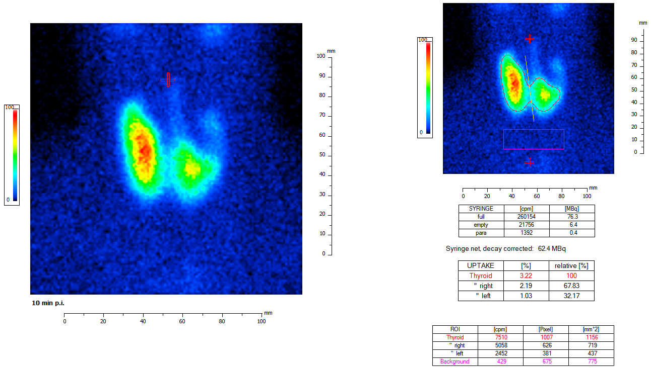
The Thyroid Program examines the function of the thyroid and parathyroid glands. It analyzes iodine uptake, assesses how the thyroid responds to hormones (suppression), and evaluates radioiodine tests to detect nodules or functional disorders. In this way, the program supports physicians in diagnosis and treatment planning.
Our Thyroid Program supports healthcare professionals in the comprehensive diagnosis of thyroid and parathyroid disorders and offers a range of specialized analysis tools:
The software enables the precise calculation of the thyroid’s relative iodine uptake relative to its overall function. In addition, the thyroid surface area and volume can be determined automatically. Three different nuclides are taken into account: Tc 99m, I 123, and I 131. The program also calculates the activity ratio of different regions of interest (ROI) to ensure a differentiated functional analysis.
The module for radioiodine tests supports evaluation for both diagnostic purposes and therapeutic dosing. The software calculates the maximum uptake or the necessary radioiodine therapy dose and performs the analysis in accordance with the guidelines of the DGN (German Society of Nuclear Medicine) and DGMP (German Society for Medical Physics).
The suppression test function allows thyroid activity to be assessed under the influence of medication. This supports the differentiated diagnosis of functional disorders.
The program offers modules for evaluating the parathyroid glands, e.g., in cases of suspected hyperparathyroidism, and facilitates the documentation of relevant parameters for treatment decisions.
Thanks to its clearly structured user interface and automated calculations, the Thyroid Program reduces the workload, increases the accuracy of diagnostic findings, and supports informed decisions in treatment planning.
The Thyroid Program is a specialized software solution for healthcare professionals designed to diagnose thyroid and parathyroid disorders.
It supports uptake calculations, radioiodine uptake (RAIU) tests, suppression tests, and parathyroid analysis, and enables automated volume and ROI calculations.
The program covers several nuclear medicine analysis methods:
The software calculates:
This enables guideline-compliant planning of radioiodine therapy.
Automated uptake calculation allows for:
This reduces manual calculation errors and significantly speeds up the workflow.
The program is intended for:
The software has been optimized for the following gamma cameras from Inter Medical:
Uptake (Iodine Uptake)
Uptake describes the percentage of radioactive iodine that the thyroid gland absorbs from the blood.
It is a key parameter for assessing thyroid function, e.g., in cases of hyperthyroidism or autonomy.
RAIU Test (Radioiodine Uptake Test)
The RAIU test measures how much radioiodine the thyroid gland has stored after a defined period of time.
It is used both for diagnosis and for calculating the radioiodine therapy dose.
ROI (Region of Interest)
An ROI is a defined area of an image in a nuclear medicine scan that is evaluated separately.
ROI analyses can be used to visualize regional functional differences within the thyroid gland or between nodules.
Suppression Test
In a suppression test, thyroid function is assessed under the influence of medication, usually after administration of thyroid hormones.
It is used to distinguish autonomously functioning areas from hormone-dependent tissue.
Hyperparathyroidism
Hyperparathyroidism is a disorder of the parathyroid glands characterized by excessive production of parathyroid hormone.
It can lead to elevated calcium levels, bone disorders, and kidney stones, and is diagnosed using nuclear medicine.
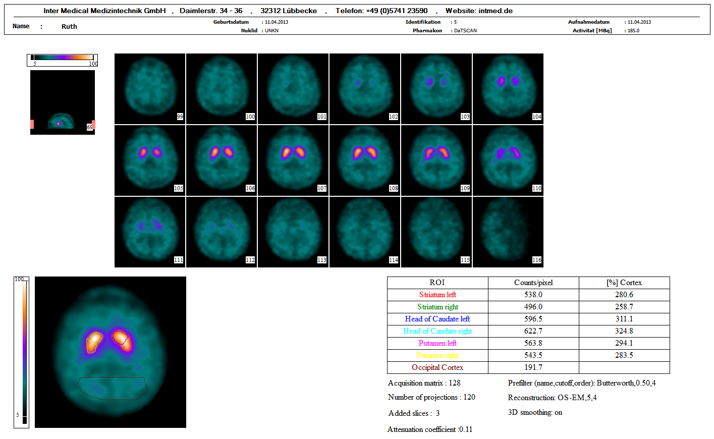
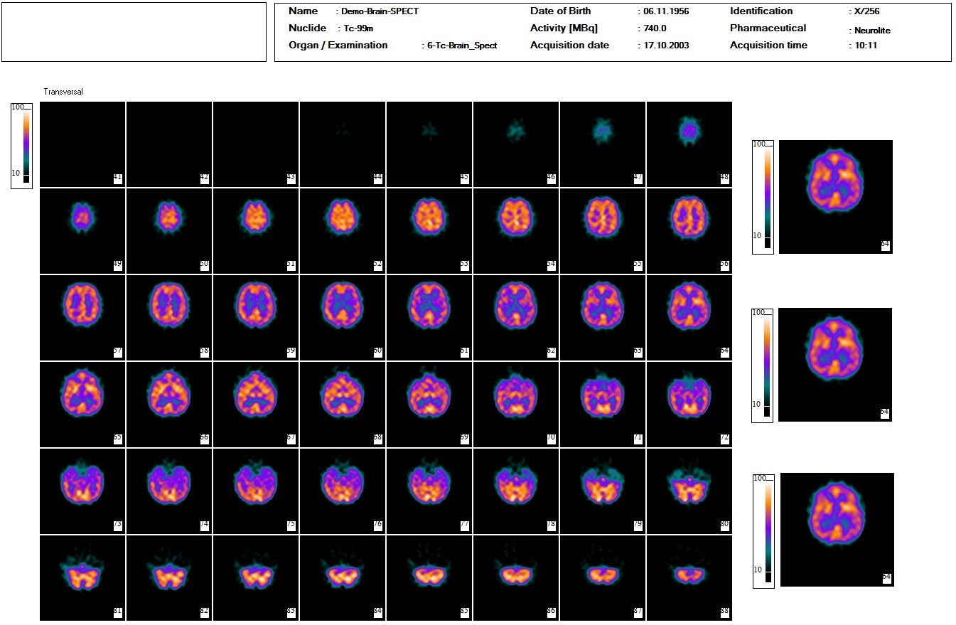
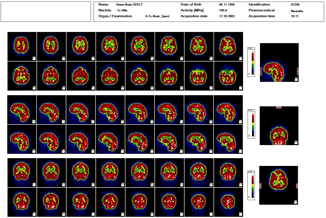
The Brain program examines the function of specific brain regions that are important for movement and the nervous system. Among other things, it analyzes DaTSCAN images to visualize dopamine transporters and IBZM scans to assess dopamine receptors. In this way, the program assists physicians in diagnosing neurological disorders such as Parkinson’s disease and other functional disorders.
Our brain program supports nuclear medicine diagnostics of the brain with specialized modules for different functional areas:
The software enables the analysis of SPECT and DaTSCAN images for the assessment of dopaminergic systems. This is particularly relevant for the diagnosis of Parkinson’s disease and other neurodegenerative disorders.
The software visualizes cerebral perfusion clearly and quantitatively, allowing for the rapid detection of circulatory disorders and functional abnormalities in the brain.
Dynamic images can be analyzed planarly, enabling precise analysis of temporal changes in tracer distribution. This supports accurate diagnoses of metabolic or circulatory disorders.
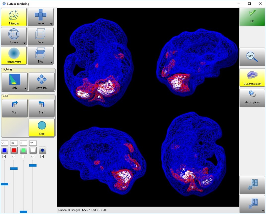
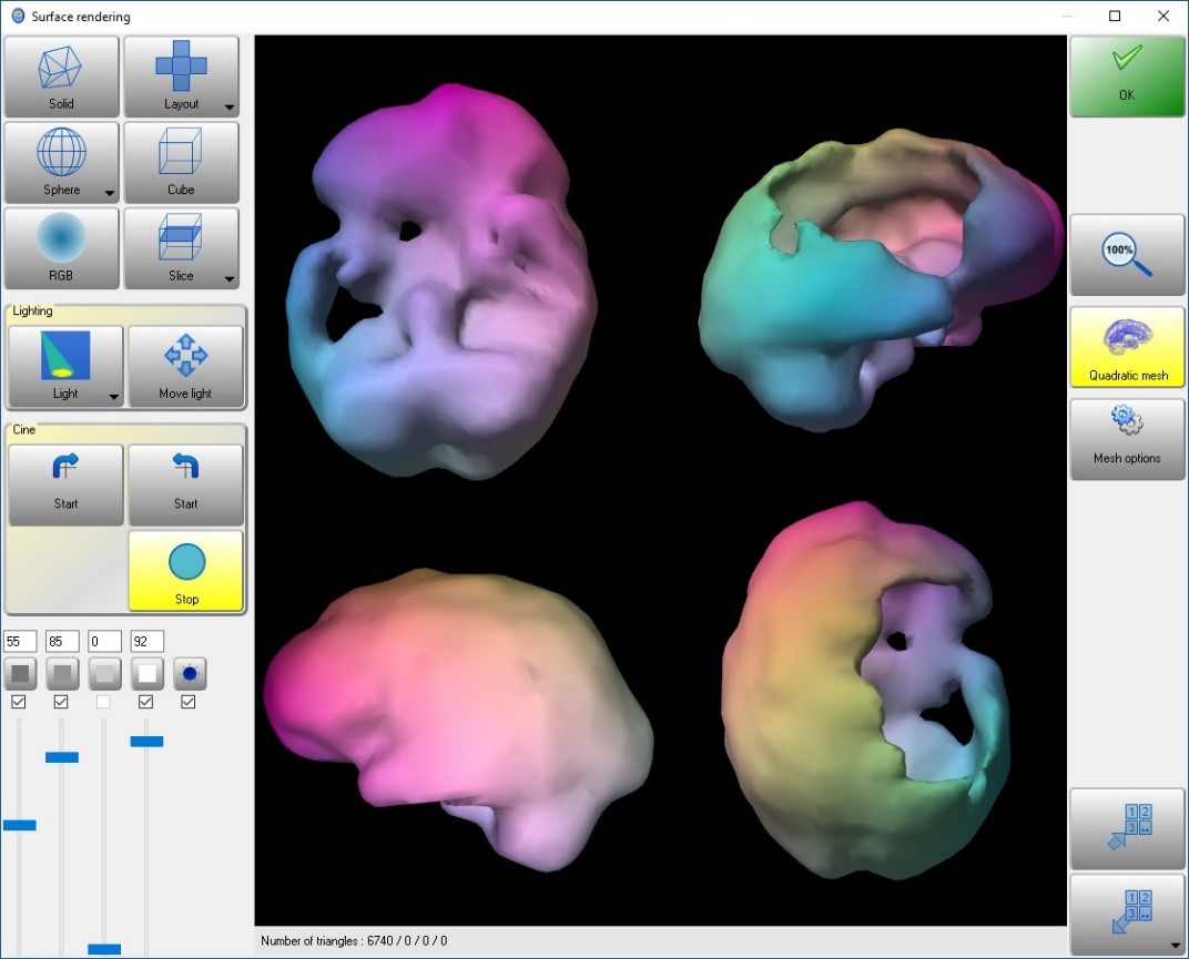
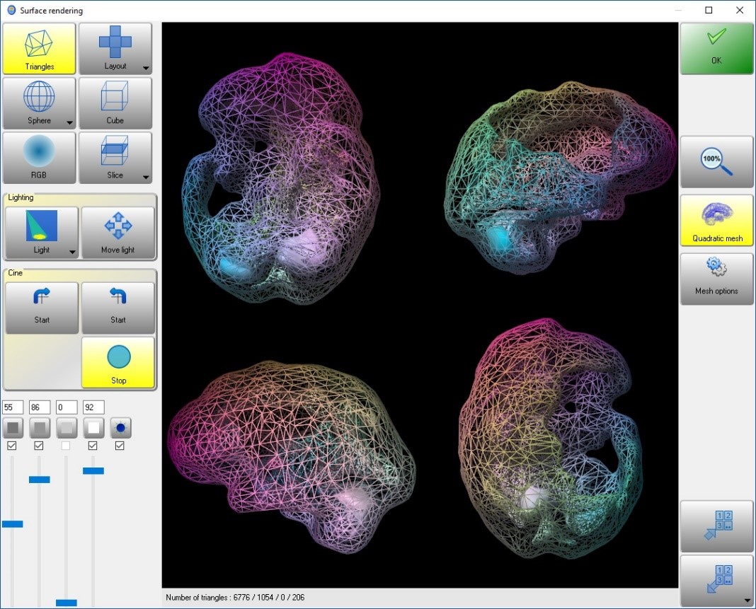
For advanced analyses, the 3D Neuro Package offers three-dimensional surface and volume visualization of cerebral perfusion. This facilitates the spatial assessment of functional disorders and supports treatment planning for complex clinical presentations.
With a clearly structured interface and automated evaluations, the brain program reduces the effort required for analysis, increases diagnostic accuracy, and provides a sound basis for decision-making in clinical practice.
The Brain Program is a specialized software solution for nuclear medical diagnostics of the brain.
It supports the evaluation of dopaminergic systems, the analysis of cerebral perfusion, dynamic examinations, and optional three-dimensional visualizations for treatment planning.
The Brain Program is used for:
The software quantitatively analyzes IBZM and DaTSCAN images to objectively assess the function of dopaminergic systems.
This is particularly relevant for differentiating between various Parkinson’s syndromes and other neurodegenerative diseases.
Cerebral perfusion imaging describes the quantitative visualization of cerebral blood flow.
The brain program clearly displays perfusion deficits and functional abnormalities, thereby facilitating diagnostic assessment.
Planar dynamic processing allows for the analysis of time-dependent changes in tracer distribution.
This enables precise assessments of circulatory or metabolic disorders and supports detailed functional diagnostics.
The 3D Neuro Package expands the analysis to include:
The Brain Program is designed for:
The software has been optimized for the following SPECT gamma cameras from Inter Medical:
DaTSCAN
DaTSCAN is a nuclear medicine examination used to visualize dopamine transporters in the striatum.
It is primarily used to diagnose Parkinson’s syndromes and to differentiate between neurodegenerative diseases.
________________________________________
IBZM
IBZM is a radiotracer used to examine postsynaptic dopamine D2 receptors.
It aids in differentiating between various forms of Parkinson’s disease and other movement disorders.
________________________________________
Cerebral Perfusion
Cerebral perfusion refers to blood flow in the brain.
Disturbances in perfusion may indicate ischemia, vascular diseases, or functional deficits and are quantitatively evaluated using nuclear medicine.
________________________________________
Planar Dynamic Imaging
Planar dynamic imaging describes the time-resolved recording of tracer distribution in a single plane.
It enables the analysis of kinetic processes in blood flow and metabolism.
________________________________________
3D Neuro Package
The 3D Neuro Package is a software module for three-dimensional visualization of cerebral perfusion. It supports volumetric analysis, surface projections, and the spatial assessment of complex neurological conditions.
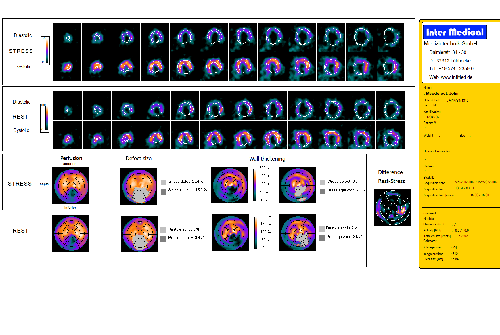
The cardiac program evaluates heart function and blood flow. It shows how well the heart is pumping, whether certain areas are damaged, and allows for comparisons between stress and rest. Automated analyses and clear visualizations help physicians identify functional abnormalities and defects with precision.
Our cardiac program offers a highly precise, automated analysis of cardiac function and perfusion and supports medical staff in diagnosis, treatment planning, and follow-up monitoring.
The software automatically recognizes cardiac structures, realigns the images, and enables a fully automated display of all diagnostically relevant cardiac parameters. This reduces manual effort and increases the accuracy of the analysis.
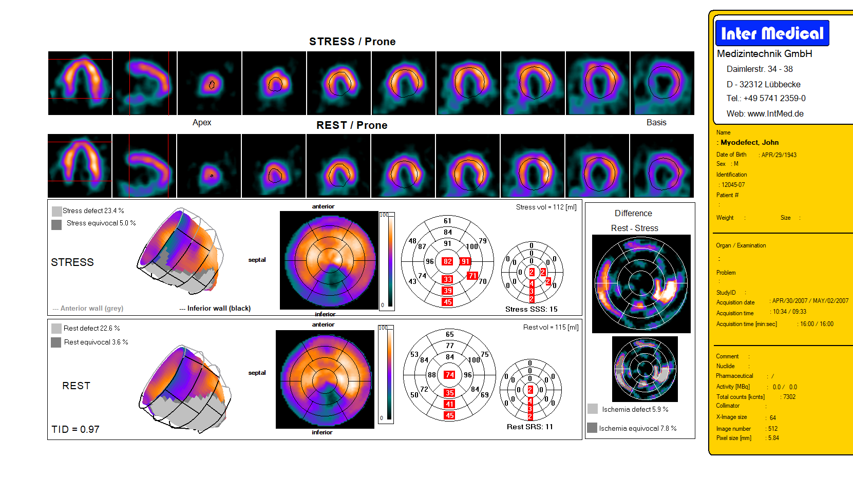
Calculation of summary scores (SSS/SRS) and defect sizes from stress and rest images—the software provides planar and cross-sectional views (HLA/VLA/short-axis) as well as polar maps for perfusion, wall thickness, wall motion, and reversibility.
These provide automatic calculation of EDV, ESV, and ejection fraction in gated studies, including volume curves, cine mode, and optional 3D ventricular visualization.
Fully standardized polar maps, in accordance with the recommendations of the American Heart Association, enable intuitive and comparable analysis.
Stress and rest images can be directly compared to identify reversible perfusion defects and functional abnormalities.
Individual patient data can be compared against a database of more than 1,000 reference patients. Additionally, motion correction (optional) and Filtered Back Projection are available for optimized image quality.
Customizable report pages, automatic display of all relevant parameters, and cross-sectional images facilitate the documentation and communication of results.
Thanks to the combination of automated processing, comparison with reference values, and detailed visualization, the cardiac program supports fast, precise, and reproducible diagnostics in cardiology and nuclear medicine.
The Heart Program is a specialized software solution for nuclear medical diagnostics of the heart.
It supports the quantitative assessment of myocardial perfusion, function, blood flow, and other cardiac parameters, and provides automated analyses for diagnosis and treatment planning.
The Heart Program is used for:
The software visualizes myocardial perfusion quantitatively and clearly.
This allows for the reliable detection of circulatory disorders, infarct scars, or regional hypoperfusion.
The cardiac program automatically determines parameters such as:
This enables physicians to assess functional disorders in detail and make well-informed treatment decisions.
Dynamic analysis enables time-resolved analysis of tracer distribution in the myocardium.
This allows for precise assessment of perfusion and metabolic processes, even during exercise or pharmacological catheterization scenarios.
The cardiac program is intended for:
The software has been optimized for these SPECT gamma cameras from Inter Medical:
Myocardial perfusion
Myocardial perfusion describes the blood flow to the heart muscle.
Disturbances in perfusion may indicate ischemia, infarct scars, or other circulatory deficits and are quantitatively assessed using nuclear medicine.
Ejection Fraction
The ejection fraction is the proportion of blood ejected from the left ventricle with each heartbeat.
It is a key parameter for assessing cardiac function.
Dynamic cardiac imaging
Dynamic cardiac imaging analyzes the temporal distribution of radiotracers in the myocardium.
This allows for the precise assessment of blood flow and metabolic processes under resting or stress conditions.
Stroke volume
Stroke volume is the amount of blood the heart ejects with each beat.
It is an important parameter for assessing cardiac output and heart health.
Coronary heart disease (CHD)
CHD is a disease of the coronary arteries characterized by reduced blood flow to the heart muscle.
It can lead to angina pectoris, heart attack, or chronic heart failure and is assessed in nuclear medicine, among other methods, using myocardial perfusion tests.
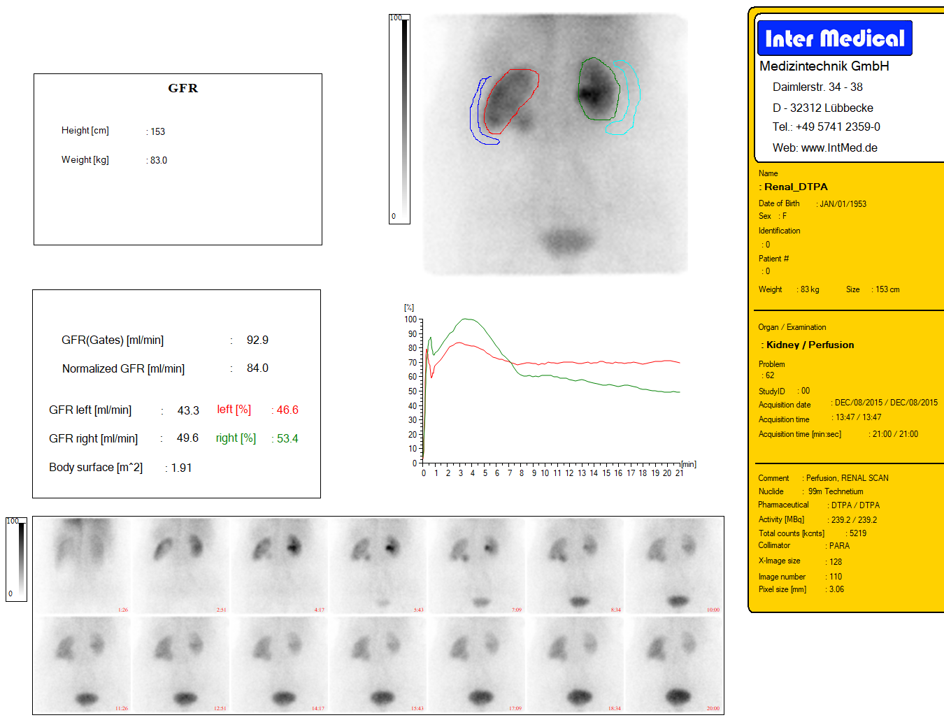
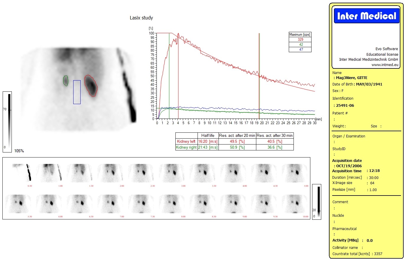
The kidney program evaluates kidney function and blood flow. It shows how well each kidney is functioning, how fluids and substances are excreted, and allows for comparisons between the two kidneys. Specialized tests, such as captopril or DMSA studies, help detect conditions such as circulatory disorders or tissue changes at an early stage.
Our kidney program supports nuclear medicine diagnostics and functional assessment of the kidneys with specialized modules for detailed evaluations:
The software enables the definition and analysis of regions of interest (ROI) for precise quantitative assessment of kidney function. Comparisons between both kidneys facilitate the identification of functional differences.
Dynamic sequence images are automatically processed and displayed in clear time-series curves to visualize functional changes over the course of the examination.
The focus here is on calculating renal clearance and the tubular excretion rate (TER) for an objective functional analysis.
This involves support for specialized tests, such as the captopril dynamic test for detecting renal artery stenosis and the DMSA scan for functional mapping of renal tissue.
With clearly structured reports, automated analyses, and intuitive graphics, the Renal Program enables efficient and precise diagnostics. This supports informed clinical decisions and simplifies the documentation of test results.
The Renal Program is a specialized software solution for nuclear medicine functional diagnostics of the kidneys.
It supports ROI analyses, clearance and TER calculations, dynamic sequence displays, and specialized examinations such as captopril and DMSA tests.
The Kidney Program is used for:
The software enables the definition and quantitative analysis of regions of interest (ROI) in both kidneys.
Comparisons of kidney function help detect asymmetric functional disorders at an early stage.
Dynamic sequence images are automatically processed and displayed as time curves.
This allows physicians to track the development of kidney function over the examination period and accurately assess changes.
Clearance: A measure of the kidney’s ability to remove substances from the blood
TER (tubular excretion rate): An assessment of tubular excretion capacity
Both parameters provide objective, quantitative data on kidney function.
Captopril-test: Identification of renal artery stenoses through pharmacological changes in blood flow
DMSA-scan: Functional mapping of renal tissue, e.g., for the detection of scarring or hypoperfusion
The kidney program is intended for:
The software has been optimized for these gamma cameras from Inter Medical:
An ROI is a defined area within an image that is specifically evaluated.
In the kidney program, it enables precise functional measurements of individual kidney segments.
Clearance indicates how efficiently the kidney removes a substance from the blood.
It is a standard parameter for assessing kidney function.
The TER describes the excretory capacity of the renal tubules.
It provides additional information on overall kidney function.
The captopril test is a nuclear medicine test used to detect renal artery stenosis.
It is based on the pharmacological change in renal blood flow following administration of captopril.
The DMSA scan uses the radiotracer dimercaptosuccinate for functional mapping of renal tissue.
It helps visualize scarring, hypoperfusion, or other structural functional deficits.
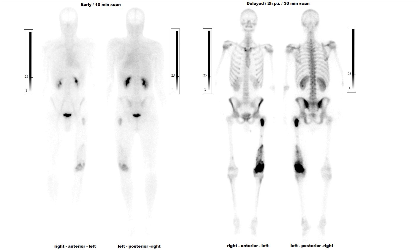
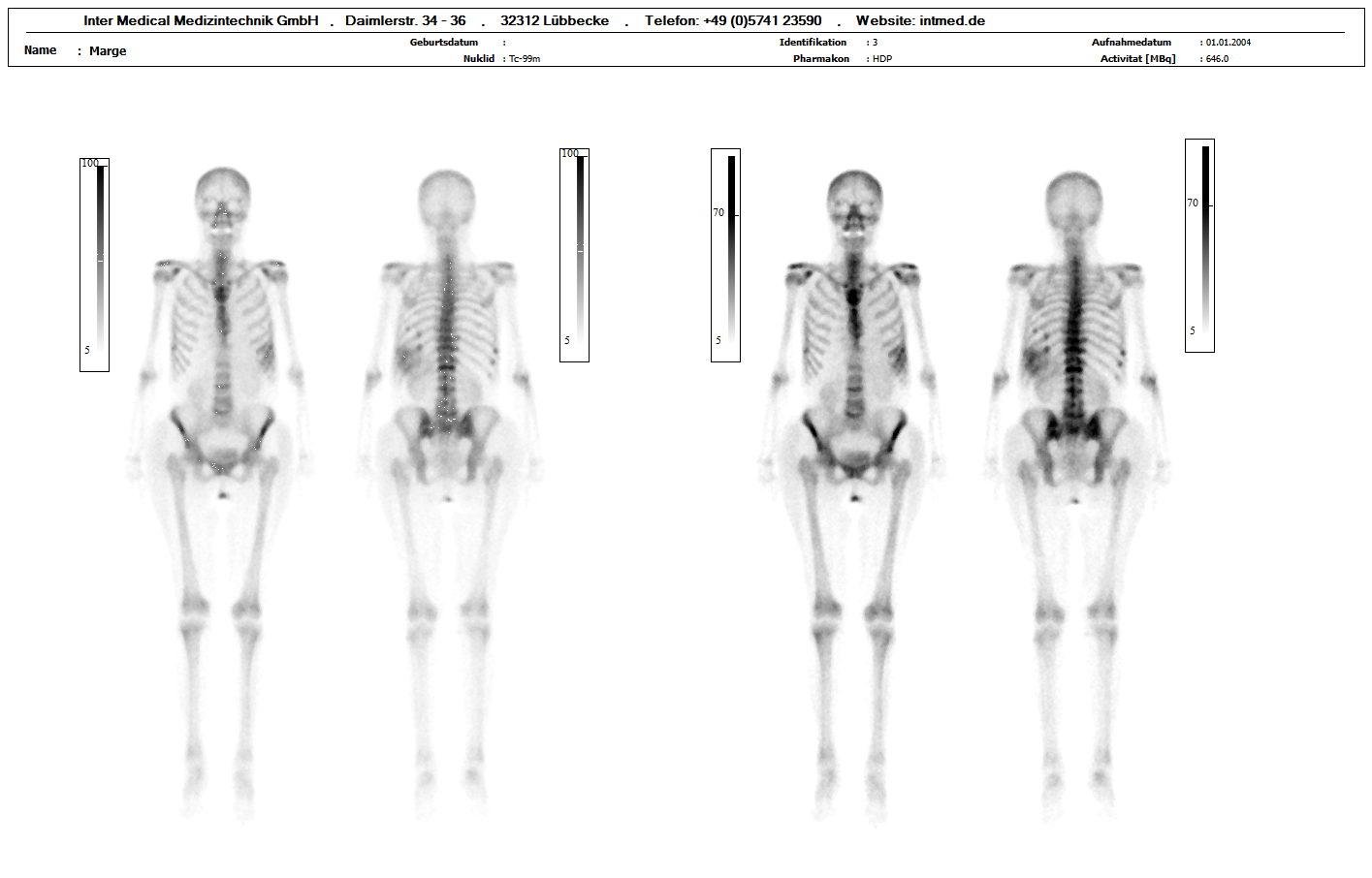
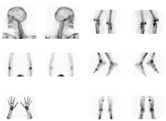
The Bone program examines the musculoskeletal and metabolic aspects of bones. It illustrates blood flow to the skeleton and the function of bone tissue from specific joints such as the sacroiliac joints to whole-body images. Dynamic images and comparisons of different regions assist physicians in diagnosing injuries, inflammation, or metabolic disorders.
Our bone program supports nuclear medicine diagnostics of the skeleton with powerful tools for quantitative and visual analysis:
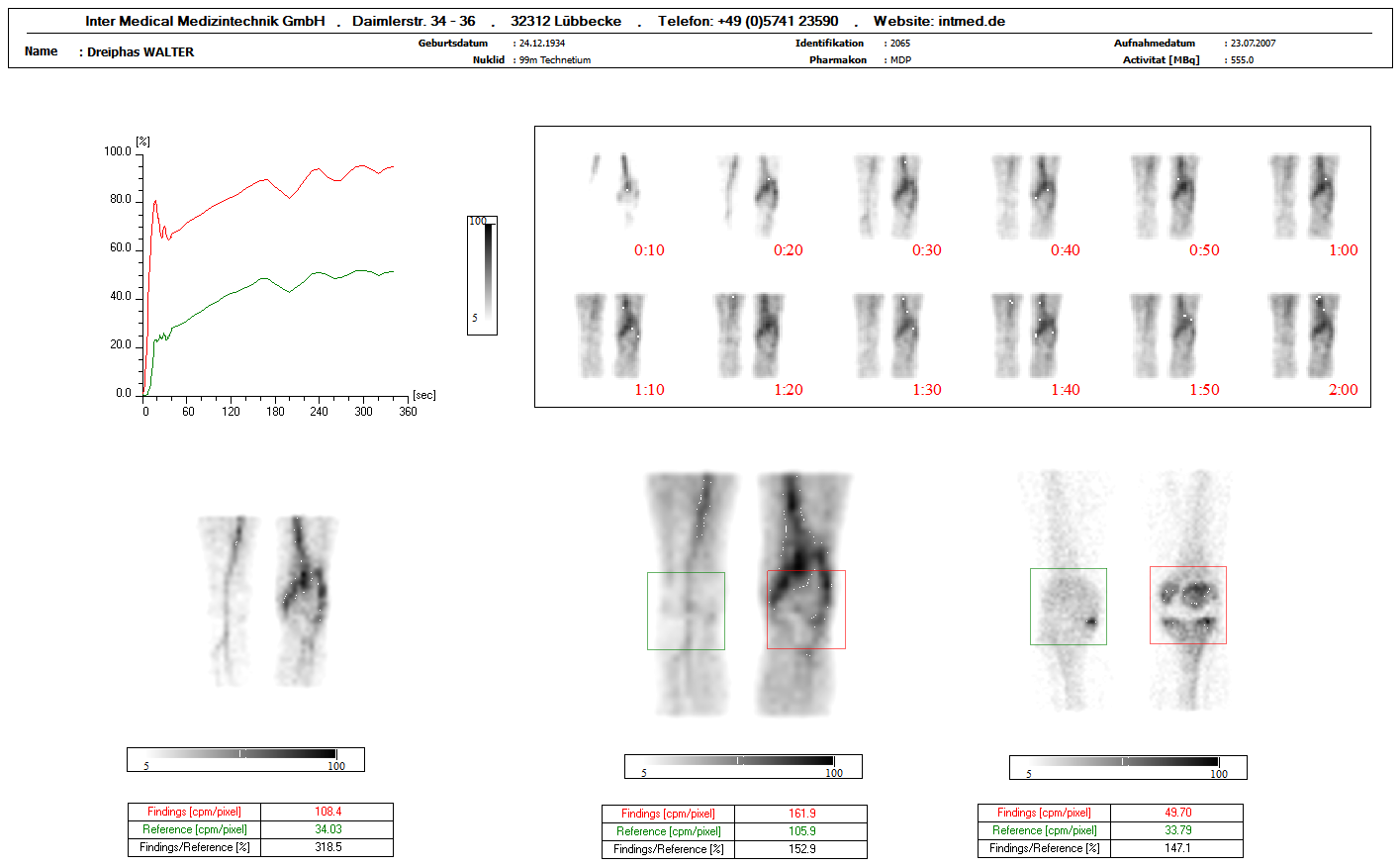
2-phase and 3-phase analyses
The software enables dynamic visualization of bone perfusion and bone metabolism via 2- or 3-phase sequences.
Sequence images and cine display
Sequence images can be displayed as curves, time series, or as cine (video) to visualize dynamic changes.
ROI analyses and comparisons
Regions of interest (ROI) can be defined and quantified across the entire skeleton. Comparisons between regions enable an objective assessment of abnormalities.
Sacroiliac joints
Specialized analysis functions for evaluating the sacroiliac joints support the diagnosis of inflammatory or degenerative changes.
Whole-body and SPECT imaging
Display of one, two, or four whole-body scans as well as SPECT images; maximum intensity projections (MIP) and flexible zoom functions facilitate the visualization of relevant areas.
Exclusion of hot areas
Areas such as injection sites or the bladder can be specifically excluded to minimize artifacts and optimize analysis.
With clear images, automated evaluations, and extensive visualization options, the Bone Program facilitates precise diagnostics, comparability of examinations, and the creation of meaningful reports.
The Bone Program is specialized software for nuclear medical diagnostics of the skeleton.
It supports 2- and 3-phase analyses, dynamic sequence displays, ROI evaluations, whole-body and SPECT visualizations, as well as specialized analyses of the sacroiliac joints.
The Bone Program is used for:
The software generates dynamic sequences of bone metabolism:
This allows for a differentiated assessment of blood flow and metabolism in specific areas.
Sequence images are automatically displayed as curves, time series, or as cine (video).
This allows time-dependent changes in bone activity to be precisely recorded and analyzed.
Regions of interest (ROI) can be specifically defined across the entire skeleton and quantitatively evaluated.
Comparisons between areas enable the objective assessment of functional differences or abnormalities.
The program offers specialized analysis tools for the sacroiliac joints to reliably identify inflammatory or degenerative changes.
Whole-body images (one, two, or four scans)
The Bone Program is intended for:
Analytical methods that dynamically depict bone metabolism in two or three time-resolved phases.
They show perfusion, blood pool, and bone uptake, enabling differentiated diagnoses.
An ROI is a defined area within the image that is specifically evaluated.
In the bone program, this allows for the quantitative analysis of individual skeletal regions.
SPECT is a nuclear medicine 3D imaging technique that visualizes the spatial distribution of radiotracers in the skeleton.
It complements whole-body or planar imaging.
The sacroiliac joints connect the sacrum and the pelvis.
Special analysis functions help detect inflammatory or degenerative changes.
MIP is a method for displaying the highest activity values within an image volume.
It facilitates the rapid identification of areas relevant to bone metabolism.
The Organ program examines key organs such as the esophagus, stomach, liver, salivary glands, lymph nodes, and eyes. It illustrates how these organs function, how quickly they transport contents or excrete fluid, and assists physicians in diagnosis and treatment planning.
Our Organ Program supports nuclear medicine diagnostics for various organ systems with specialized modules for quantitative and visual analysis:
Esophagus
Calculation of transit time and analysis of esophageal channel function to precisely identify motility disorders
Stomach
Processing and analysis of gastric scintigraphy to assess gastric emptying, motility, and functional disorders
Liver (HIDA)
Analysis of liver and bile duct function, including calculation of the gallbladder ejection fraction (EF), for the objective assessment of hepatobiliary diseases
Salivary glands
Quantitative and visual evaluation of salivary gland function, e.g., in cases of reduced secretion or inflammatory processes
Sentinel lymph nodes (SLN)
Support in the identification and processing of sentinel lymph nodes for preoperative planning and diagnosis
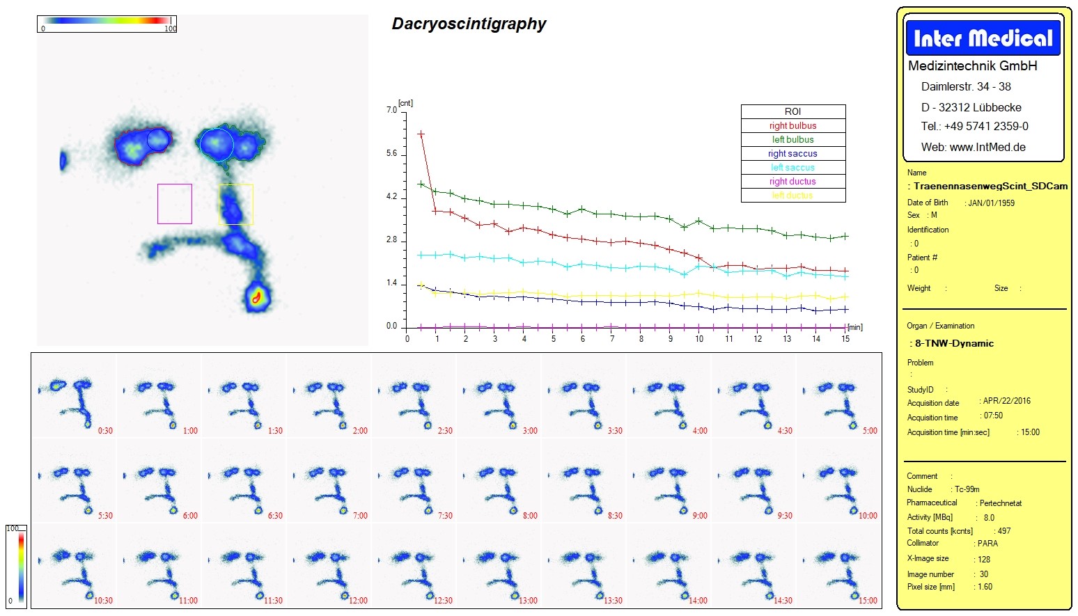
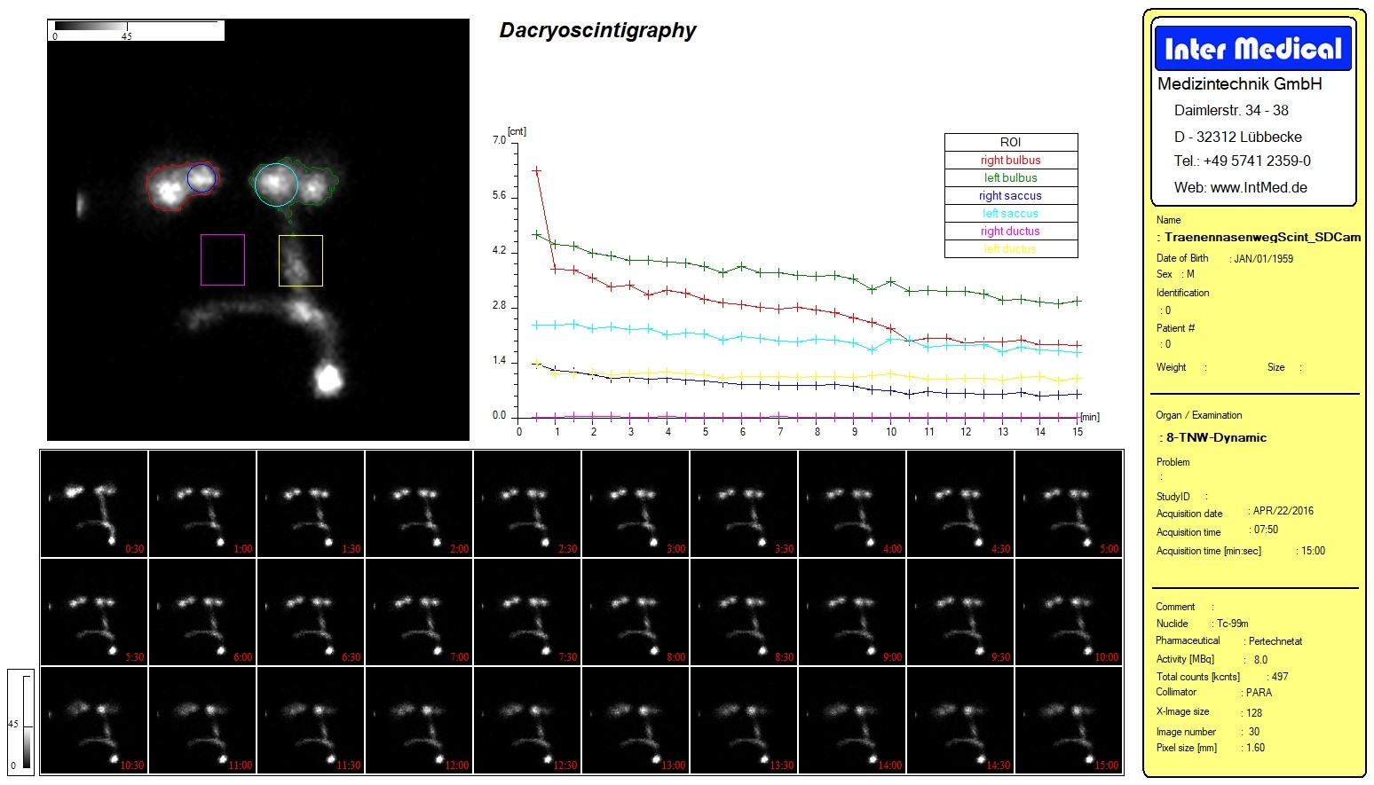
Eyes (dacryoscintigraphy)
Analysis of lacrimal duct function for the diagnosis of dacryostenosis or secretory disorders
With automated analyses, graphical representations of time series, and standardized calculations, the Organ Program enables efficient, precise, and reproducible diagnostics of various organ systems.
The Organ Program is a specialized software solution for nuclear medicine diagnostics of multiple organ systems.
It supports quantitative analyses, visualizations, and automated calculations for the esophagus, stomach, liver, salivary glands, sentinel lymph nodes, and eyes.
The Organ Program is used for:
The software calculates transit time and evaluates the function of the esophageal channel.
This allows motility disorders to be objectively detected and documented.
Gastric scintigraphy is analyzed quantitatively and visually, including:
This enables precise diagnosis of gastrointestinal problems.
The HIDA evaluation analyzes liver and gallbladder function, including calculation of the gallbladder ejection fraction (EF).
This allows for an objective assessment of hepatobiliary diseases.
Salivary glands: quantitative and visual functional analysis
Sentinel lymph nodes (SLN): identification and preoperative planning
Eyes (dacryoscintigraphy): analysis of lacrimal duct function
The Organ Program is intended for:
Transit time describes the time it takes for a bolus to pass through the esophagus.
It is an important parameter for assessing esophageal motility disorders.
Gastric scintigraphy is a nuclear medicine examination used to assess gastric emptying, motility, and functional disorders.
HIDA is a nuclear medicine examination used to assess liver and gallbladder function.
The ejection fraction (EF) of the gallbladder can be quantitatively determined.
The sentinel lymph node is the first lymph node to which a tumor typically metastasizes.
Identifying it aids in preoperative planning and the diagnosis of oncological diseases.
Dacryoscintigraphy is a nuclear medicine procedure used to examine tear duct function.
It is used to diagnose dacryostenosis or secretory disorders.
👷 This content is still under development!
👷 This content is still under development!
👷 This content is still under development!
Analysis modules for standards requirements
👷 This content is still under construction!
👷 This content is still under construction!